-
CDST_LT: Introduction to MGIT 960 system
FullscreenMGIT 960 Instrument: Operation
ContentThe MGIT 960 is a sensitive instrument to be operated by trained and competent staff. To carry out the liquid culture using this instrument, following steps need to be performed.
Table: Operation of the MGIT 960 Instrument
Step 1
To load the tubes, open the desired drawer.
Once the drawer is open, select the desired workflow on the LCD display, in this case, Tube Entry.

Step 2
Scan pre-affixed barcode on tube
Step 3
Load where indicated by solid green LED light in drawer, and close the drawer.
Now the MGIT 960 system will do all the work.
Step 4
Wait until the MGIT 960 system either flags the tube as positive or negative. After opening the drawer, remove the positives and completed negatives as they occur (icons appear on each drawer) eg press positive that appears on the screen to pull out positive tube from the slot.
Resources
Kindly provide your valuable feedback on the page to the link provided HERE
-
CDST_LT: Sample processing reagents preparation
FullscreenPrecautions for Sample Processing and Reagent Preparation in TB Culture Labs
ContentPrecautions for sample processing and reagent preparation in TB culture labs are as follows:
- Precaution is necessary while performing aerosol-generating procedures such as centrifugation, vertexing, mixing, pipetting, pouring and inoculation of media. For example -delay the opening of caps until aerosols have settled, open centrifuge canisters only inside the Bio Safety Cabinets (BSCs), use pipettes that are easy to control.
- Safety precautions to minimize interruption of airflow inside the BSC by keeping arms parallel to the work surface inside the BSC and working in the center to minimize arm movements and ensure not to move hands out of the hood until work is completed. Minimize the equipment inside the BSC so that there is no interference in the airflow pattern.
- Disinfect the BSC and all work surfaces with a tuberculocidal disinfectant before and after every procedure.
- The autoclave should be monitored with a spore test, at least monthly, to ensure that sterility is achieved.
- Avoid practices that can result in spills.
- Train all personnel working in the TB culture lab.
- Working staff must wear Personal Protective Equipment (PPE) like protective gowns, gloves, hair covers and shoe covers etc.
Resources
Kindly provide your valuable feedback on the page to the link provided HERE
BACTEC MGIT 960 Growth Supplement
ContentThe MGIT 960 Growth Supplement (Figure below) is the enrichment added to the MGIT medium prior to inoculation of specimen.
It provides substances essential to the growth of mycobacteria, such as:
- Oleic acid, that is utilized by tubercle bacteria and plays an important role in the metabolism of mycobacteria.
- Albumin, that acts as a protective agent by binding free fatty acids which may be toxic to Mycobacterium species, thereby enhancing their recovery.
- Dextrose, that is an energy source.
- Catalase, that destroys toxic peroxides that may be present in the medium.
- Polyoxyethylene stearate, that enhances the growth of Mycobacterium tuberculosis and assists in providing a uniform inoculum.
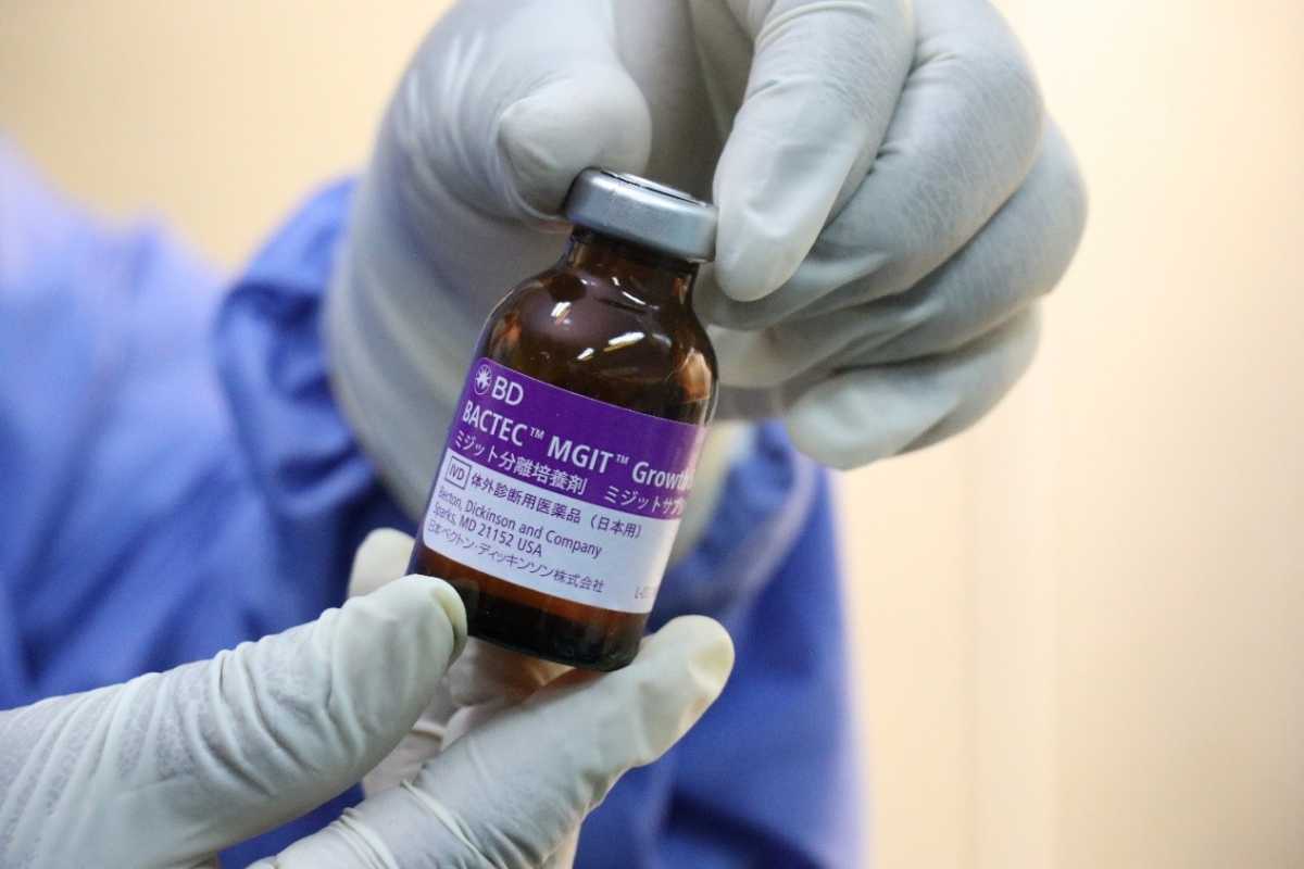
Figure: MGIT 960 Growth Supplement
Constituents of MGIT 960 Growth Supplement
15 ml MGIT 960 Growth Supplement contains:
- Bovine Albumin: 50.0 gm
- Dextrose: 20.0 gm
- Catalase: 0.03 gm
- Oleic Acid: 0.1 gm
- Polyoxyethylene state (POES): 1.1 gm
NOTE: It is a sterile product, do not use if turbid or contaminated.
Resources
Kindly provide your valuable feedback on the page to the link provided HERE
Quality Control of MGIT 960 Tubes
ContentMGIT 960 media quality control (QC) is to be done upon receipt of a new shipment or lot number. This includes checking the media visually and by growing control strains of Mycobacterium. Steps include:
- Visually check the tube for:
- Turbidity/contamination
- Expiry date
- Breakage
- Transport condition
- Control organisms prepared in Middlebrook 7H9 Broth should be used for QC testing as per the procedure shown in the figure below. It is important to test all three organisms, as each have different requirements for growth, and each will demonstrate a different time to detection. A good quality control program ensures that the media supports the growth of a variety of organisms, not just one organism.
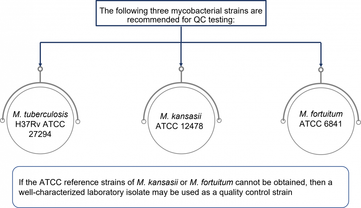
Figure: Control strains used for quality control of MGIT tubes
- Quality control testing should be performed in a biosafety cabinet
-
Subculture the quality control strains on Lowenstein-Jensen (LJ) slants and use pure and fresh growth to prepare a uniform suspension of 0.5 McFarland turbidity
- Dilution of culture suspension using sterile saline or distilled water, accordingly:
- M. tuberculosis - 1:500
- M. fortuitum - 1:5000
- M. kansasii - 1:50000
- Inoculate 0.5 ml of respective dilutions of culture suspension into two labeled MGIT tubes and further incubate the tubes into MGIT 960 system
- The expected results are:
- M. tuberculosis: Positive in 6 to 10 days
- M. kansasii: Positive in 7 to 11 days
- M. fortuitum: Positive in 1 to 3 days
Resources
Kindly provide your valuable feedback on the page to the link provided HERE
- Visually check the tube for:
-
CDST_LT: Specimen processing and inoculation
FullscreenSpecimen Processing for TB Cultures
ContentSputum specimens are viscous materials contaminated with normal flora. Therefore, processing involves pre-treatment of the sputum specimens via:
- Digestion: To free the TB bacilli from the mucus in which they may be embedded.
- Decontamination: To eradicate normal flora that grows more rapidly than TB bacilli, and would interfere with the ability to recover TB bacilli.
- Homogenization: Of the digested materials.
- Concentration: Of the TB bacilli by centrifugation before smear preparation and media inoculation.
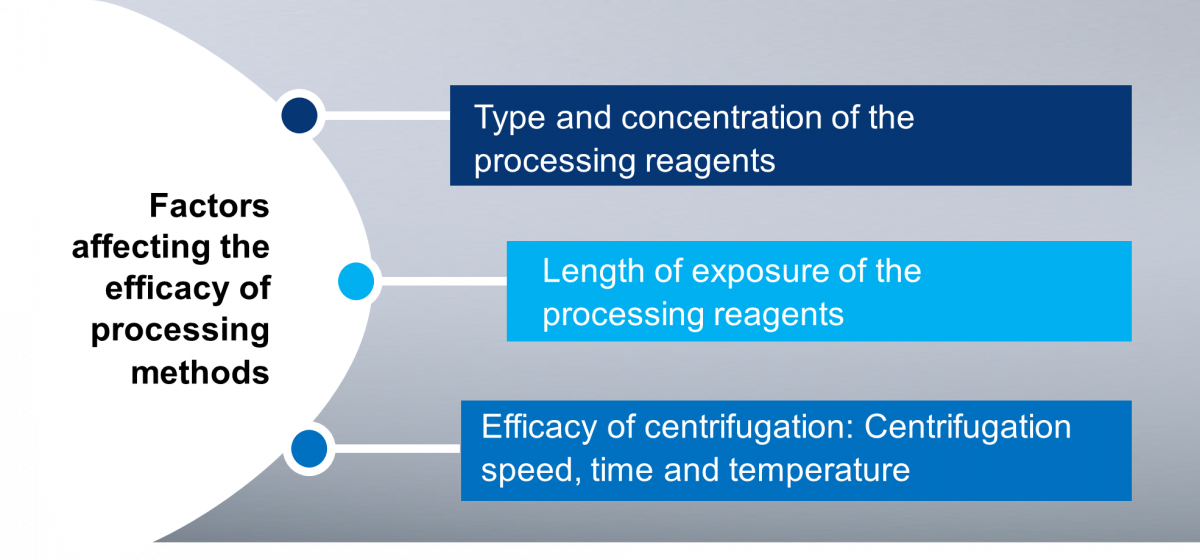
Figure: Factors Affecting the Efficacy of Processing Methods
Methods of Culture Specimen Processing
Various processing methods are used for TB specimens; amongst them, the most common methods are:
- N-acetyl-L-cysteine - sodium hydroxide (NALC-NaOH) method: It is the mildest decontamination method which can kill about 33% of mycobacteria in a clinical specimen. It can be used with both liquid and solid media.
- Petroff’s sodium hydroxide method: It is a harsher method – it can kill up to 70% of mycobacteria in specimens. Although useful with highly contaminated specimens, it is not recommended for use with liquid MGIT media.
Resources
- MGIT Procedure Manual, Mycobacteria Growth Indicator Tube (MGIT) Culture and Drug Susceptibility Demonstration Projects, FIND Training Manual.
- GLI Training Module on Specimen Processing, STOP TB Partnership.
Kindly provide your valuable feedback on the page to the link provided HERE
Procedure for Culture Specimen Processing: Pulmonary specimens
ContentThese are the steps to be followed when processing pulmonary specimens in TB culture laboratories:
Beginning the Specimen Processing Procedure:
- Process only one specimen at a time, and do not leave open containers or open centrifuge tubes in the Bio Safety Cabinet (BSC).
- Process the available specimen in a 50 ml sterile, plastic, screw-capped centrifuge tube (Figure).
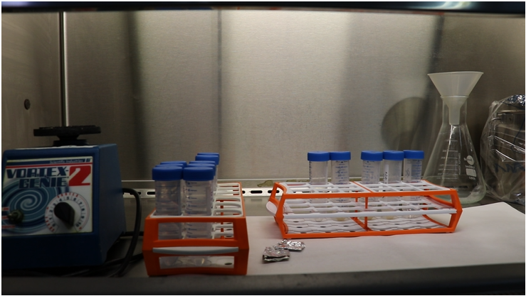
Figure: Capped 50 ml sterile, plastic, screw-capped centrifuge tubes
NALC - NaOH Procedure:
- Always open the cap of the specimen container slowly to minimize aerosol production.
- Aliquot reagent in a separate tube for each specimen to avoid contamination of reagent stocks. A freshly prepared single-use aliquot is preferred.
- Note the volume of the specimen. Add an equal volume of N-acetyl-L-cysteine-sodium hydroxide (NALC-NaOH) solution and tighten the cap.
- In order to avoid cross-contamination, do not allow the NALC-NAOH solution container to touch the specimen tubes.
- After the addition of the decontaminant and or digestant tighten the caps and vortex for not more than 20 seconds at a moderate speed.
- Invert each tube 5 times, making the figure 8 with your wrist, to ensure that the NALC-NaOH solution contacts the entire inner surface of the tube and cap.
- Avoid extreme agitation or shaking which can cause inactivation of the NALC.
- Let the tubes stand at room temperature for 20-25°C, for 15 minutes and mix by gently inverting the tube.
- NaOH exposure time must be strictly limited to 15 minutes to prevent the over-killing of the TB bacilli.
- If stronger decontamination is needed, the starting concentration of NaOH may be increased to 5-6%, but the time of exposure should not be extended.
- Neutralize the specimen with a phosphate buffer of pH 6.8 to the 45 ml mark. Do not exceed the 45 ml mark.
- To avoid cross-contamination do not allow the diluent (phosphate buffer) container to touch the mouth of the specimen tubes.
- Single-use aliquots of phosphate buffer or a dispenser are preferred to avoid cross-contamination during the procedure.
- After centrifugation, open the safety bucket in the BSC and carefully pour off the supernatant into a splash-proof discard container with a suitable disinfectant (5% phenol).
- If required, swab the tube with a disinfectant-soaked gauze (use individual pieces) and recap carefully.
- While wiping, do not allow the disinfectant to flow into the tube.
- Re-suspend the sediment in 1–2 ml of sterile phosphate buffer (pH 6.8) using a new transfer pipette.
Please click here to see a full video on sputum specimen processing for culture.
Resources
- MGIT Procedure Manual, Mycobacteria Growth Indicator Tube (MGIT) Culture and Drug Susceptibility Demonstration Projects, FIND Training Manual.
- GLI Training Module on Specimen Processing, STOP TB Partnership.
Kindly provide your valuable feedback on the page to the link provided HERE
Procedure for Culture Specimen Processing: Extrapulmonary specimens
ContentExtrapulmonary specimens are divided into 2 groups based on the site and mode of collection and the extent of contamination:
- Aseptically collected specimens, usually free from other microorganisms (sterile), which include fluids like spinal, pleural, pericardial, synovial, ascitic, blood, bone marrow, tissues (lymph node, tissue biopsies) and fine needle aspirates (FNAs)
- Specimens contaminated by normal flora or specimens not collected aseptically (not sterile), such as gastric lavage, bronchial washings, urine, pus, other muco-purulent specimens and stool (in case of disseminated TB in HIV infected patients and infants)
All extrapulmonary specimens have to be appropriately collected, transported, registered, decontaminated, cultured via solid culture methods or processed for MGIT960.
All extrapulmonary specimens have different processing procedures that need to be used for the respective specimen.
Resources
Kindly provide your valuable feedback on the page to the link provided HERE
Procedure for Culture Specimen Processing: Pus and other muco-purulent specimens
ContentThe procedure for processing pus and other muco-purulent specimens in TB cultures is as follows:
- If the specimen is thick or mucoid and less than 10 ml in volume, digest and decontaminate with N-acetyl-L-cysteine-sodium hydroxide (NALC-NaOH) method similar to the procedure used for sputum specimens.
- If the specimen is not thick, it may be treated with 2-4% NaOH.
- The concentration of NaOH depends upon the contaminating bacteria expected to be present in the specimen.
- If the volume is over 10-12 ml, process only 10 ml or the first concentrate by centrifugation at 3000x g for 15-20 minutes.
- In such a situation, if the specimen is thick, liquefy the specimen by adding a small quantity of NALC only (50-100 mg powder) and mix well.
- After the concentration step, resuspend the sediment in 5 ml sterile water, decontaminate with NaOH and concentrate again by centrifugation.
- Always resuspend the sediment (pellet) in buffer to reduce the pH.
Resources
Kindly provide your valuable feedback on the page to the link provided HERE
Procedure for Culture Specimen Processing: Gastric Aspirate
ContentThe procedure for processing gastric aspirates in TB cultures is as follows:
- Concentrate by centrifugation before decontaminating.
- Resuspend the sediment in about 5 ml of sterile water and decontaminate with N-acetyl-L-cysteine-sodium hydroxide (NALC-NaOH) method.
- After decontamination, concentrate again prior to inoculation of the sediment into the culture media.
- Due to the low pH, gastric aspirates should be processed as soon as possible (within 4 hours of collection).
- If the specimen cannot be processed quickly, it should be neutralized with NaOH before transportation or storage.
Resources
Kindly provide your valuable feedback on the page to the link provided HERE
Procedure for Culture Specimen Processing: Bronchial Lavage
ContentThe procedure for processing bronchial lavage specimens in TB cultures is the same as that for sputum samples. The following aspects need to be considered:
- If the specimen is larger than 10 ml in volume, it needs to be concentrated by centrifugation (3000x g, 15-20 minutes) and the sediment resuspended in 5 ml sterile water.
- Thick/ mucoid samples need to be liquefied by mixing the specimen with a pinch (50-100 mg) of N-acetyl-L-cysteine (NALC) powder.
The processed specimen is used to inoculate the MGIT tubes.
Resources
Kindly provide your valuable feedback on the page to the link provided HERE
Procedure for Culture Specimen Processing: Laryngeal Swab
ContentThe procedure for processing laryngeal swab specimens in TB cultures is as follows:
- Transfer the swab (without a stick) into a sterile centrifuge tube and add 2 ml sterile water.
- Add 2 ml N-acetyl L-cysteine - sodium hydroxide (NALC-NaOH) solution, replace the cap, mix well (vortex mix), keep for 15 minutes.
- Remove the swab with forceps, squeezing the liquid out of the swab, and discard the swab.
- Fill the tube with phosphate buffer, mix and centrifuge at 3000x - 3500x g (15-20 minutes).
- Discard the supernatant fluid and resuspend the sediment in 1-2 ml sterile buffer.
The processed specimen is used to inoculate the MGIT tubes.
Resources
Kindly provide your valuable feedback on the page to the link provided HERE
Procedure for Culture Specimen Processing: Tissue
ContentThe procedure for processing tissue specimens in TB cultures is as follows:
- Add saline or water (2 - 4 ml) and homogenize the tissue in a Biosafety Cabinet (BSC) using sterile equipment:
- Tissue grinder or homogenizer
- If these are not available, use a mortar and pestle
- Small tissue specimens may be placed in a petri dish with 2-4 ml sterile water and torn apart with the help of two sterile needles
- Decontaminate the homogenized specimen with N-acetyl L-cysteine - sodium hydroxide (NALC-NaOH) procedure as in processing sputum. Tissue biopsies collected aseptically do not require decontamination procedures.
- Resuspend the sediment in phosphate buffer.
The processed specimen is used to inoculate the MGIT tubes.
Resources
Kindly provide your valuable feedback on the page to the link provided HERE
- Add saline or water (2 - 4 ml) and homogenize the tissue in a Biosafety Cabinet (BSC) using sterile equipment:
Procedure for Culture Specimen Processing: Urine
ContentThe procedure for processing urine in TB cultures is as follows:
- Aliquot the entire volume in several centrifuge tubes.
- Concentrate the specimen by centrifugation in 50 ml centrifuge tubes for 20-25 minutes.
- Resuspend the pellet in each tube with 1-2 ml of sterile water and pool together with a total volume of 5-10 ml.
- Decontaminate sediment with 4% sodium hydroxide (NaOH) for 15-20 minutes and proceed as similar to processing sputum.
The processed specimen is used to inoculate the MGIT tubes.
NOTE: Isolation of mycobacteria from urine specimens using MGIT has not been validated by Becton, Dickinson & Company (MGIT 960 manufacturer) but other investigators have reported successful isolation of mycobacteria from urine specimens. When performed, the sample to be used is:
- Totally voided, early morning urine specimen
- Pooled or mid-stream urine is not recommended.
Resources
Kindly provide your valuable feedback on the page to the link provided HERE
Procedure for Culture Specimen Processing: CSF and other body fluids
ContentBody fluids collected aseptically (cerebrospinal fluid, synovial fluid, pleural fluid) can be inoculated into MGIT medium without decontamination (with the addition of PANTA). However, since it is difficult to maintain sterile conditions throughout the collection of specimens, it is recommended that all specimens be decontaminated.
Aseptically collected specimens need only light decontamination (NALC without NaOH). Steps for processing such specimens are:
- Large volume samples (>10 ml) are concentrated by centrifugation at 3000x - 3500x g (15-20 minutes).
- N-acetyl-L-cysteine (NALC) powder (50-100 mg) is added to thick/ mucoid specimens prior to centrifugation.
- Resuspend the sediment in 5 ml saline.
- Decontaminate as per the procedure for sputum samples.
The processed specimen is used to inoculate the MGIT tubes.
Resources
Kindly provide your valuable feedback on the page to the link provided HERE
Steps to be done after Culture Specimen Processing
ContentAfter specimen processing, the resuspended pellet may be used for various tests as per the laboratory policy or national guidelines recommended for TB diagnosis such as:
- Inoculation of sediments in Mycobacterium Growth Indicator Tube (MGIT) broth (Liquid media) and or solid media for primary culture isolation
- Subculture a loopful on a Brain Heart Infusion (BHI) Agar plate for contamination check
- Concentrated smear of processed specimen can be prepared if not prepared earlier, for a microscopy smear
- DNA extraction for Line Probe Assay
Transfer the remaining sediment into 2 mL labelled cryovials and store at 2-8⁰C for repeat or additional testing.
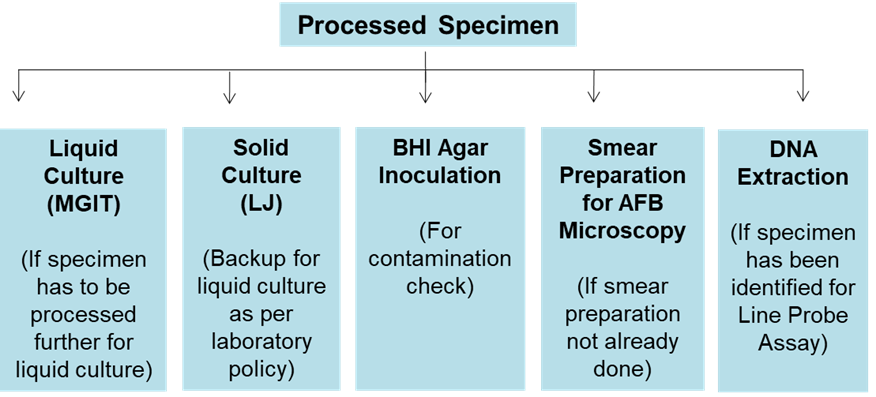
Figure: Steps After Sample Processing
Resources
Kindly provide your valuable feedback on the page to the link provided HERE
Preparation of PANTA for MGIT TB Cultures
ContentPreparation of PANTA is the first step when inoculating and incubating MGIT 960 tubes for TB cultures. The preparation steps are shown below (Figure 1).

Figure 1: Steps for Preparation of PANTA

Figure 2: Reconstitution of PANTA with growth supplement using a micropipette
NOTE:
- Reconstituted PANTA can be used for up to 5 days if stored at 2 to 8ºC, but do not freeze.
- Each PANTA vial is used for 15-18 MGIT tubes (BACTEC MGIT 960).
Please click the video below to see the full procedure for inoculating and incubating MGIT tubes for TB cultures:
Resources
Kindly provide your valuable feedback on the page to the link provided HERE
Preparation of MGIT Tubes for MGIT TB Cultures
ContentPreparation of MGIT tubes occurs after preparation of PANTA when inoculating and incubating MGIT 960 tubes for TB cultures. The preparation steps are shown below.
- Prepare the MGIT tubes in a clean Biosafety Cabinet (BSC) preferably prior to specimen processing.
- Put an absorbent sheet on the work surface of the BSC and soak it with disinfectant (bactericidal for mycobacteria).
- Before preparation of MGIT tubes:
- Know the number of samples for batch or per day
- Label each 7 ml MGIT tube with a specimen number
- Decontaminate all items to be used like micropipettes, racks, tip boxes and vortex
- Aseptically, add 0.8 ml of PANTA mixture to each MGIT tube.
- Inoculate tubes with specimen within 2 hours of adding PANTA.
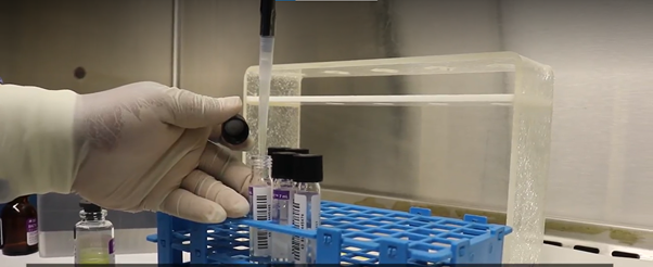
Figure: Addition of 0.8 ml of PANTA/ Growth supplement mixture to the MGIT tube using a micropipette
Please click the video below to see the full procedure for inoculating and incubating MGIT tubes for TB cultures:
Resources
Kindly provide your valuable feedback on the page to the link provided HERE
Inoculation of MGIT Tubes
ContentMycobacteria Growth Indicator Tube (MGIT) contains a modified Middlebrook 7H9 broth base. When supplemented with MGIT Growth Supplement and PANTA, it provides an optimum medium for the growth of a majority of mycobacterial species. All types of specimens, pulmonary as well as extra-pulmonary (except blood), can be inoculated into MGIT for primary isolation of mycobacteria.
The steps for inoculating MGIT tubes are elaborated below:
- Using a sterile transfer pipette, add 0.5 ml of the processed sample to a 7 ml MGIT tube (0.8 ml of PANTA-growth supplement mixture already added).
- Tightly recap the tube and invert gently several times to mix well.
- Recap and swab the exterior of each tube with disinfectant-soaked gauze (use individual swabs); ensure the disinfectant does not flow into the tube.
- Leave the inoculated tubes at room temperature for 30 minutes.
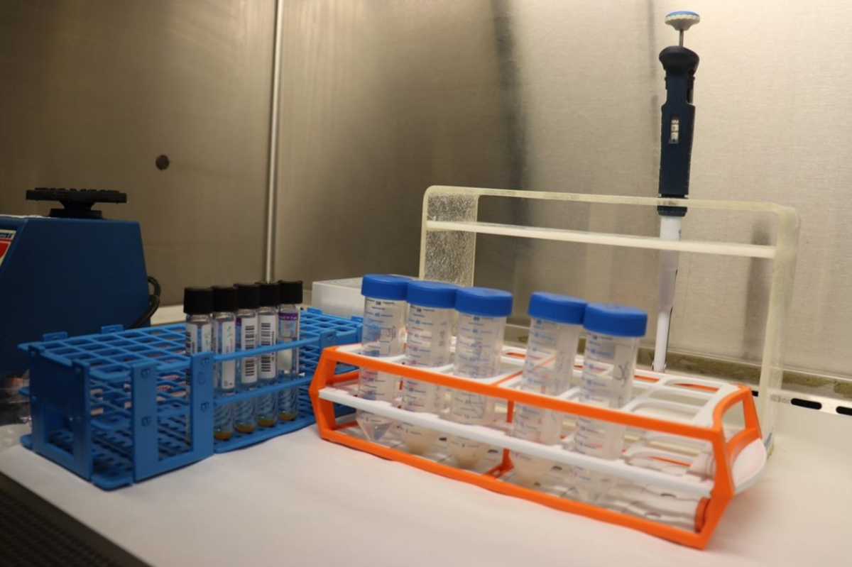
Figure: Micropipette that will be used to add processed sputum specimen to the prepared MGIT tube
Precautions
- Do not add more than 0.5 ml of processed specimen.
- Volumes greater than 0.5 ml may alter the pH of the medium and result in false-positive fluorescence.
- Use a separate pipette or pipette tip for each specimen.
- Always recap the tube tightly, as loose caps may affect the detection of fluorescence.
Please click the video below to see the full procedure for inoculating and incubating MGIT tubes for TB cultures:
Resources
Kindly provide your valuable feedback on the page to the link provided HERE
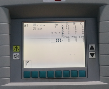
Incubation of MGIT Tubes
ContentAfter inoculating the tubes, they have to be incubated in the BACTEC MGIT 960 instrument to check if there is mycobacterial growth after the stipulated time.
The steps for incubating MGIT tubes for TB cultures are as follows:
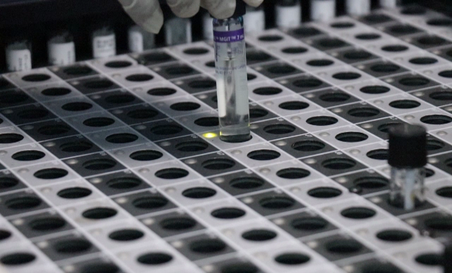
1. Enter the inoculated MGIT tubes into BACTEC MGIT 960 instrument.
2. Scan the tube bar code and specimen bar code (if available) (Figure 1).
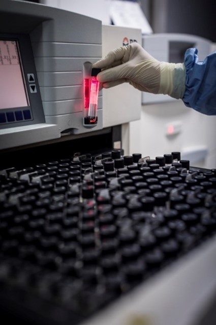
Figure 1: Scanning the tube bar code
3. Insert tubes into the instrument in the slots assigned (indicated by a green light) (Figure 2).

Figure 2: Slots with green light indicated for inserting the tubes
4. The tubes are incubated in the MGIT 960 instrument at 37±1°C.
a. The temperature readout of each drawer must be checked daily.
b. Since the optimum temperature for growth of M. tuberculosis is 37ºC, make sure the temperature is close to 37ºC.
5. Fluorescence in each tube is measured every 60 minutes by the photodetector assembly.
6. Tubes are detected Positive or Negative, based on the fluorescence.
7. MGIT tubes should be incubated until the instrument flags them positive. After a maximum of six weeks, the instrument flags the tubes negative if there is no growth.
8. Some species such as M. ulcerans and M. genavense may require extended incubation time; if such species are expected to be present, incubate further for 2-3 weeks.
Please click the video below to see the full procedure for inoculating and incubating MGIT tubes for TB culture:
Resources
Kindly provide your valuable feedback on the page to the link provided HERE
-
CDST_LT: Growth detection
FullscreenBACTEC MGIT 960 System Growth Detection
ContentBACTEC MGIT 960 System Growth Detection
Principle:
The MGIT (Mycobacteria Growth Indicator Tube) consists of a liquid broth medium in the form of Middlebrook 7H9 liquid media that is known to yield better recovery and faster growth of mycobacteria. The MGIT tube also contains an oxygen-quenched fluorochrome, tris 4, 7-diphenyl-1, 10-phenanthroline ruthenium chloride pentahydrate embedded in silicone at the bottom of the tube.
During bacterial growth within the tube, the free oxygen is utilized and is replaced with carbon dioxide. With the depletion of free oxygen, the fluorochrome is no longer inhibited, resulting in fluorescence within the MGIT tube when visualized under UV light. The intensity of fluorescence is directly proportional to the extent of oxygen depletion. MGIT tubes may be incubated at 37ºC and read manually under a UV light or entered into an MGIT 960 instrument, where they are incubated and monitored for increasing fluorescence every 60 minutes.
The growth of bacteria, as well as mycobacteria, increases the fluorescence. In the case of M. tuberculosis, at the time of positivity, there are approximately 105 – 106 colony-forming units (CFU) per ml of medium. The instrument declares a tube negative if it remains negative for six weeks (42 days). The detection of growth can also be visually observed by the presence of non-homogeneous light turbidity or a small granular/flaky appearance in the medium.
Notifications:
Positive Cultures:
The system will indicate the presence of presumptive positive vials in several ways:
-
The positive indicator lamp at the front of the drawer illuminates.
-
The tube count for each drawer, next to the filled circle with a plus sign icon, increments in the Summary window display.
-
When the drawer is opened, the "remove positive tubes" soft key appears on the screen.
-
The audible alert sounds until the condition is acknowledged.
Negative Cultures:
Negative cultures exist as ongoing negatives (in the protocol) or out-of-protocol negatives. Notification of these conditions includes:
-
Ongoing Negatives - In the summary region of the display, the ongoing tube count for each drawer appears next to the filled circle icon,
-
Out-of-protocol Negatives - In the summary region of the display, the tube count for each drawer appears next to the filled circle with a minus (- )sign icon.
-
The negative indicator light for the drawer(s) illuminates.
Resources
1. MGIT Procedure manual, FIND Diagnostics
Assessment
Question 1
Answer 1
Answer 2
Answer 3
Answer 4
Correct Answer
Correct Explanation
Page id
Part of Pre-Test
Part of Post-Test
The MGIT (Mycobacteria Growth Indicator Tube) consists of a liquid broth medium in the form of Middlebrook 7H10 liquid media.
True
False
False
The MGIT (Mycobacteria Growth Indicator Tube) consists of a liquid broth medium in the form of Middlebrook 7H9 liquid media that is known to yield better recovery and faster growth of mycobacteria.
Yes
Yes
Question 2
The instrument declares a tube negative if it remains negative for six weeks (46 days).
True
False
False
The instrument declares a tube negative if it remains negative for six weeks (42 days).
Yes
Yes
-
Work-up for Positive MGIT Cultures
ContentThe different steps of tube removal are as follows:
1. Visual Observation:
Characteristic flake-like growth (Presumptive Positive)
Uniform Turbidity (Presumptive Contamination)
2. Smear:
Corded AFB smear-positive (Culture positive)
Both Acid Fast or Non-Acid Fast Bacilli (Culture with contamination)
Only Non-Acid Fast Bacilli (Contamination)3. Inoculate LJ slant/BHI for contamination check by the rate of growth, morphology, smear and immunochromatographic assays
Growth in 24 – 48 hours (Contamination)
No Growth (Contamination free)
Cultures should be checked for purity and identification of M. tuberculosis using a rapid immunochromatographic species identification test, such as MPT 64 Ag Assay
Resource
Mycobacteriology Laboratory Manual
Assessment
Question 1
Answer 1
Answer 2
Answer 3
Answer 4
Correct Answer
Correct Explanation
Page id
Part of Pre-Test
Part of Post-Test
AFB smear should be prepared from a positive MGIT tube.
True
False
True
AFB smear should be prepared from a positive MGIT tube.
Yes
Yes
Question 2
Sub-culturing a positive MGIT tube on an LJ slant is recommended.
True
False
True
Sub-culturing a positive MGIT tube on an LJ slant is recommended.
Yes
Yes
Interpreting MGIT Results
ContentMGIT results are interpreted as described in Flowcharts 1-3:
Image Image
Image Image
Image
Resource
Mycobacteriology Laboratory Manual
Assessment
Answer 1
Answer 2
Ans
wer 3
Answer 4
Correct answer
Correct explanation
Page id
Part of Pre-test
Part of Post-test
How is early growth in the MGIT tube interpreted? Re-incubate for 42 days Sub-culture MGIT broth on LJ slant Rapid ID test from MGIT All 4 Flowchart 3 is followed to interpret early growth in MGIT, which includes re-incubation for 42 days, sub-culturing MGIT broth on LJ slant and rapid ID test from MGIT.
Yes Yes
-
CDST_LT: Liquid culture contamination
FullscreenLiquid Culture Contamination and sources of contamination
ContentLiquid Culture Contamination and Sources of Contamination
Liquid media are more prone to contamination than solid media. Therefore, it is essential to process specimens with extreme care, adhering very closely to procedures and recommendations.
Sources of Contamination:
- Improper or under-decontamination of the specimen
- Thick mucoid specimens that are hard to liquefy
- Prolonged storage and transportation time of the specimen after collection. In such situations, especially in hot weather, bacteria tend to overgrow and are hard to kill by routine decontamination procedures.
-
Use of non-sterile materials such as pipettes, tubes, etc.
The incidence of contamination with bacteria (other than mycobacteria) varies from laboratory to laboratory, depending upon several factors. According to the NTEP guidelines, up to a 5% contamination rate is acceptable in cultures of clinical specimens on solid media. However, for liquid media, slightly higher contamination may be accepted (up to 7-8%). A very low contamination rate (less than 3%) may indicate too harsh a decontamination process, which would also affect the growth of mycobacteria and may reduce the positivity rate and increase the time-to-detection of positive mycobacterial culture.
Resource
- Assessment
Question 1
Answer 1
Answer 2
Answer 3
Answer 4
Correct Answer
Correct Explanation
Page id
Part of Pre-Test
Part of Post-Test
The acceptable range of contamination in solid culture is up to 5%.
True
False
True
According to the NTEP guidelines, up to a 5% contamination rate is acceptable in cultures of clinical specimens on solid media.
Yes
Yes
Question 2
The acceptable range of contamination in liquid culture is up to 7-8%.
True
False
True
According to the NTEP guidelines, up to 7-8% contamination rate is acceptable in cultures of clinical specimens on liquid media.
Yes
Yes
- Improper or under-decontamination of the specimen
Monitoring Liquid Culture Contamination
ContentThe growth of contaminant bacteria results in positive fluorescence. Therefore, it is important to observe all fluorescent positive MGIT tubes visually for turbidity and to make an AFB smear. If an MGIT tube broth is heavily turbid, contamination is suspected even if the AFB smear is positive.
Usually, contaminating bacteria causes heavy turbidity, although M. tuberculosis growth appears as particles without significant turbidity, while some of the NTM may produce light turbidity.
Contamination may be confirmed by the following method:
1. Make a smear and stain with Ziehl-Neelsen stain. The presence of non-acid-fast contaminant bacteria on the smear confirms contamination.
2. Sub-culture a loopful on blood agar. If blood agar is not available, use chocolate agar or a brain heart infusion (BHI) agar plate. Several specimens (up to 4) may be carefully inoculated on a plate (small streak for each specimen, properly labelled). Divide the plate and identify the specimen number with a marker. Incubate these subcultures at 35ºC ±1ºC and observe after 24-48 hours. If contaminating growth appears, confirm again by gram and ZN stain.
3. If contamination is confirmed with a negative AFB smear from the broth, discard the specimen and report it as contaminated. The isolation procedure can be repeated if contamination is confirmed with a positive AFB smear from the broth.
Resource
Mycobacteriology Laboratory Manual
Assessment
Question 1
Answer 1
Answer 2
Answer 3
Answer 4
Correct Answer
Correct Explanation
Page id
Part of Pre-Test
Part of Post-Test
Liquid culture contamination can be monitored by inoculating on blood agar.
True
False
True
Sub-culture can be done on blood agar. If blood agar is not available, chocolate agar or brain heart infusion (BHI) plate may be used.
Yes
Yes
Question 2
If contamination is confirmed with a negative AFB smear from the broth, discard the specimen and report it as contaminated.
True
False
True
If contamination is confirmed with a negative AFB smear from the broth sample, it should be discarded and reported as contaminated.
Yes
Yes
-
CDST_LT: False culture results
FullscreenFalse Positive MGIT Cultures
ContentFalse positives are test results reported as positive for a Mycobacterium species not present in the patient specimen.
-
Not all false positives are due to laboratory cross-contamination.
-
All laboratories are capable of producing false positive results.
Practices that can lead to false positive cultures are:
-
Inadequate sterilization of instruments such as bronchoscopes
-
Mislabeling at the time of collection or time of accessioning
-
Use of contaminated water for specimen collection or laboratory procedures
-
Shared reagents and dispensers
-
Opening more than one specimen container at a time while processing
-
Mix-up of testing samples or lids
-
Failure to take precautions to minimize aerosol production
Resource
Assessment
Question 1
Answer 1
Answer 2
Answer 3
Answer 4
Correct Answer
Correct Explanation
Page id
Part of Pre-Test
Part of Post-Test
False positives are test results reported as positive for a Mycobacterium species not present in the patient specimen.
True
False
True
False positives are test results reported as positive for a Mycobacterium species not present in the patient specimen.
Yes
Yes
Question 2
The use of contaminated water for specimen collection or laboratory procedures is one of the reasons for false positives.
True
False
True
Among several reasons, the use of contaminated water for specimen collection or laboratory procedures is one of the reasons for false positives.
Yes
Yes
-
False Negative MGIT Cultures
ContentFalse negatives are test results reported as negative for a Mycobacterium species present in the patient specimen
Practices that can lead to false negative cultures are :
-
Extended exposure to NaOH during processing
-
High pH (insufficient neutralization during processing)
-
Growth may be inhibited by:
-
addition of too much PANTA
-
loss of CO2 from MGIT vial headspace
-
Resource
DocumentMGIT 960 Growth Detection Oct09.ppt (1.46 MB)Assessment
Question 1
Answer 1
Answer 2
Answer 3
Answer 4
Correct Answer
Correct Explanation
Page id
Part of Pre-Test
Part of Post-Test
False negatives are test results reported as negative for a Mycobacterium species present in the patient specimen.
True
False
True
False negatives are test results reported as negative for a Mycobacterium species present in the patient specimen.
Yes
Yes
Question 2
Extended exposure to NaOH during processing results in false negatives.
True
False
True
Extended exposure to NaOH during processing results in false negatives.
Yes
Yes
-
Impact of False MGIT Culture Results
ContentLaboratories need a mechanism to determine possible causes of false results (e.g., personnel logs, lot numbers used). Practices should be evaluated to determine where changes can be made. Most episodes of false results are recognized only after a review of laboratory records, including genotyping results. Laboratories need a review process to detect false cultures earlier.
Consequences of false positives which impact on clinical care of patients:
1. Patients may be managed incorrectly in the following ways.
-
Unnecessary treatment and toxicity
-
Unnecessary isolation, hospitalization, and healthcare costs
-
Emotional repercussions to the patient
-
Unnecessary contact investigations
2. Credibility of the laboratory, hospital, or clinician may be questioned.
3. It May increase laboratory workload and testing costs.
Resource
Assessment
Question 1
Answer 1
Answer 2
Answer 3
Answer 4
Correct Answer
Correct Explanation
Page id
Part of Pre-Test
Part of Post-Test
False positive results may lead to unnecessary treatment and toxicity.
True
False
True
False positive results may cause unnecessary treatment of the patient and cause toxicity.
Yes
Yes
Question 2
Due to false positive results credibility of the laboratory, hospital, or clinician may be questioned.
True
False
True
False positive results affect the credibility of the laboratory, hospital, or clinician.
Yes
Yes
-
-
CDST_LT: Liquid culture troubleshooting
FullscreenTroubleshooting LC Growth Recovery
ContentIf there is an overall decrease in the culture positivity rate, the following parameters need to be investigated:
A. Incubation:
The majority of mycobacterial species grow well at 37°C ± 1ºC. They may grow slowly or may not grow if the temperature drops below 35°C. If an incubator is used, confirm that the incubator's temperature is 37°C ± 1°C by placing a calibrated thermometer in various locations throughout the incubator or instrument drawers. Monitor the readings several times each day until heating stability is determined. Check the temperature of the MGIT 960 by retrieving the information from the instrument.
B. Decontamination Procedure:
-
Confirm that the purity and concentration of all the reagents used in the digestion/ decontamination procedure are satisfactory.
-
Use distilled/ deionized water only for the preparation of reagents.
-
It is better to start with freshly prepared reagents.
-
A high pH of the specimen inoculated into the MGlT medium may influence the performance of MGlT adversely.
-
Do not expose the specimen to the decontamination reagent for longer than the recommended time.
- Check if MGlT tubes are positive by visual growth but negative by fluorescence.
C. Centrifugation:
-
Relative Centrifugal Force (RCF) should be 3,000-5,000 x g. Make sure the centrifuge is giving the required RCF.
-
The generation of heat during centrifugation also lowers the recovery due to higher temperature. Avoid the generation of excessive heat by using a refrigerated centrifuge.
D. Use of PANTA:
Check that PANTA is reconstituted with the proper volume.
Resource
Mycobacteriology Laboratory Manual
Assessment
Question 1
Answer 1
Answer 2
Answer 3
Answer 4
Correct Answer
Correct Explanation
Page id
Part of Pre-Test
Part of Post-Test
The majority of mycobacterial species grow well at 37°C + 1ºC.
True
False
True
Most mycobacterial species grow well at 37°C + 1ºC; however, some may require temperatures other than 37°C.
Yes
Yes
Question 2
The reagents for decontamination can be used, which were prepared 1 month back.
True
False
False
It is better to use freshly prepared reagents.
Yes
Yes
-
Troubleshooting LC Detection Time
ContentDetection time can range from 24 hours to six weeks for MGIT and 8 weeks for LJ medium. If the average detection time is significantly longer, these instructions need to be followed:
Digestion/decontamination procedure:
-
Decrease the NaOH concentration and/or time of exposure to NaOH. Higher concentrations of NaOH or longer exposure time will prolong the detection time of mycobacteria.
-
A high pH of the final inoculum will prolong the detection time.
-
Check if the incubation temperature is within specifications. Lower temperatures would delay detection.
-
In a few instances, too high a concentration of PANTA may delay the detection of certain strains of NTM, especially if the starting number is low.
Procedures Check:
-
Water used to prepare reagents should be pure (distilled/deionized).
-
All the reagents used should be sterile.
-
All pipettes and tubes should be sterile.
-
All inoculations should be made in the biological safety hood.
-
Growth Supplement/PANTA mixture should be added to MGIT tubes just before inoculation.
-
The addition of MGlT OADC or MGIT Growth Supplement/PANTA must be done inside a biological cabinet. Leave the tube open for as little time as possible. Leaving the tube open, especially on an open bench top, would increase the contamination rate.
Resource
Mycobacteriology Laboratory Manual
Assessment
Question 1
Answer 1
Answer 2
Answer 3
Answer 4
Correct Answer
Correct Explanation
Page id
Part of Pre-Test
Part of Post-Test
Higher concentrations of NaOH or longer exposure time will prolong the detection time of mycobacteria.
True
False
True
Higher concentrations of NaOH or longer exposure time kill a lot of Mycobacteria.
Yes
Yes
Question 2
Growth Supplement/PANTA mixture can be added to MGIT tubes after inoculation.
True
False
False
Growth Supplement/PANTA mixture should be added to MGIT tubes just before inoculation.
Yes
Yes
-
Troubleshooting High Liquid Culture contamination
ContentA high contamination rate indicates improper decontamination procedure. At the same time, too low contamination indicates over-treatment of the specimen that could also lower the culture positivity rate or increase the detection time. If in the MGIT it is more than 7-8%, then the decontamination procedure is not satisfactory and corrective measures should be taken:
A. Specimen collection and transport:
1. Collect specimens in clean and sterile containers to avoid outside contamination.
2. Keep the specimen in cool conditions during transport, preferably in an insulated ice box.
3. Transport to the lab as quickly as possible.
4. Upon receipt, keep it in a cool place, preferably in a refrigerator.
5. Process the specimen as soon as possible.
B. Specimen quality and quantity:
1. The sample should not be too watery or too mucoid. If a mucoid specimen is not completely liquefied, add a small quantity of NALC powder.
2. The volume of the digested and decontaminated specimen should be 2.0–10.0 ml.
C. Specimen processing:
1. NALC-NaOH is the method of choice.
2. Recommended NaOH concentration of 4% is ideal (the final concentration in the specimen is 1%).
3. An increase in NaOH usually lowers the contamination rate.
4. Higher NaOH concentration (up to 1.5% in the specimen) is acceptable when contamination is a serious problem. Once the contamination problem is under control, try to lower the NaOH concentration gradually and bring it to the recommended concentration.
D. Addition of PANTA:
1. Check storage conditions and expiry date of lyophilized PANTA (refrigerated at 2- 8ºC). Improper preparation or storage of PANTA can affect the performance or optimal concentrations.
2. Once reconstituted can be stored at 2-8ºC within 5 days and may not be frozen.
E. Specimen inoculation:
-
The specimen should be inoculated inside a safety cabinet.
-
Tubes should be inoculated with the correct amount (0.5 ml) of the specimen.
-
The inoculated MGIT tubes should be mixed after adding the PANTA and specimen.
Quality control:
-
Process 5 ml sterile buffer (negative control) along with a regular batch of specimens processed in a day. Process the negative control in the same way as clinical specimens and inoculate them into MGIT tubes. This would indicate if there is a source of contamination during the processing.
-
Periodic sterility testing of the reagents, especially a freshly made batch, is required to keep a check on the contamination sources from the reagents. Use a blood agar plate or any other suitable bacteria medium for checking contamination and Middlebrook agar or LJ medium for mycobacterial contamination check.
-
Environmental contamination may be reduced by thoroughly disinfecting the lab, working inside a biosafety hood for all the additions and other processes, and fixing the source of contamination, if established.
Resource
Mycobacteriology Laboratory Manual
Assessment:
Question 1
Answer 1
Answer 2
Answer 3
Answer 4
Correct Answer
Correct Explanation
Page id
Part of Pre-Test
Part of Post-Test
The specimen can be inoculated outside the cabinet.
True
False
False
The specimen should be inoculated inside the cabinet. Otherwise, the chances of contamination will increase.
Yes
Yes
Question 2
Quality control measures should be followed at regular intervals for liquid culture.
True
True
True
Quality control measures should be followed at regular intervals for liquid culture.
Yes
Yes
-
Decontamination of Contaminated MGIT Culture
ContentUsually, more than one specimen is collected from a patient. Therefore, it is not necessary to salvage a contaminated specimen if other specimens from the same patient are positive and not contaminated. However, if it is critical to have the results of a particular specimen that was contaminated, the broth may be reprocessed.
The steps involved are as follows:
-
The entire contaminated MGIT broth should be transferred into a 50 ml centrifuge tube.
-
An equal quantity of sterile 4% sterile NaOH solution.
-
This should be mixed well and allowed to stand for 15-20 minutes while mixing the tube by inverting it periodically.
-
Phosphate buffer of pH 6.8 should be added after 15-20 mins up to the 40 ml mark on the centrifuge tube and mixed well by inverting the tube.
-
Centrifugation should be done at 3000g for 15-20 minutes.
-
The supernatant fluid is poured off.
-
The sediment is resuspended in 0.5ml phosphate buffer (6.8 pH) and mixed well.
-
0.5 ml of this is inoculated into a fresh MGIT tube supplemented with the MGIT growth supplement/PANTA.
-
The inoculated tubes are left at room temperature for 30 minutes and then placed in the instrument. This is followed up for observation of growth.
-
If no growth is observed, Autoclave all inoculated MGIT tubes before disposal.
Resource
Mycobacteriology Laboratory Manual
Assessment
Question 1
Answer 1
Answer 2
Answer 3
Answer 4
Correct Answer
Correct Explanation
Page id
Part of Pre-Test
Part of Post-Test
Contaminated culture can be decontaminated.
True
False
True
if it is critical to have results of a particular specimen that was contaminated, the broth may be reprocessed.
Yes
Yes
Question 2
The sediment is resuspended in 0.8 ml of phosphate buffer (6.8 pH).
True
False
False
The sediment is resuspended in 0.5ml of phosphate buffer (6.8 pH).
Yes
Yes
-
-
CDST_LT: MGIT Reading and reporting
FullscreenDocumentation of MGIT Cultures
ContentDocumentation of MGIT cultures is important to characterise if growth is positive for M. tuberculosis or unexpected result/contamination. The growth is tested for AFB by ZN microscopy and sterility by inoculation on the BHI agar/LJ slant.
The documentation of growth is as follows:
MGIT Machine Result
Growth Characteristics
ZN Result
BHI agar/LJ slant result
Documentation
Positive
Flakes like growth
Positive
No growth
M. tuberculosis complex detected
Positive
Flakes like growth
Plus turbidity
Positive
Growth
M. tuberculosis complex detected
with contamination
Positive
Flakes like growth
Positive
No Growth
M. tuberculosis complex not detected but positive for NTMs
Positive
Turbidity
Negative
Not Applicable
Contamination
Negative
No growth
Not Applicable
Not Applicable
Negative for M. tuberculosis complex
Assessment
Question 1
Answer 1
Answer 2
Answer 3
Answer 4
Correct Answer
Correct Explanation
Page id
Part of Pre-Test
Part of Post-Test
Flaky and turbid positive MGIT growth indicates which of the following?
M. tuberculosis complex with contamination
M. tuberculosis complex
M. tuberculosis complex not detected
NTM detected
1
Flaky and turbid positive MGIT growth indicates M. tuberculosis complex with contamination.
Yes
Yes
Question 2
Flakes like growth, MGIT positive but does not grow on BHI/LJ indicates which of the following?
M. tuberculosis complex with contamination
M. tuberculosis complex
M. tuberculosis complex not detected but positive for NTMs
E. coli detected
3
Flakes like growth, MGIT positive but does not grow on BHI/LJ indicates M. tuberculosis complex not detected but positive for NTMs.
Yes
Yes
MGIT Culture Reading Schedule
Content-
In automated systems, tubes are read continuously and flagged when positive. Performing acid-fast bacteria (AFB) smear with Zeihl-Neelsen (ZN) staining determines the next steps.
-
All MGIT-negative tubes at the end of the incubation period should be visually checked for evidence of growth before being discarded.
-
ZN staining should be performed on growth as soon as possible. After staining, culture should be handled according to the results.
Resource
Assessment
Question 1
Answer 1
Answer 2
Answer 3
Answer 4
Correct Answer
Correct Explanation
Page id
Part of Pre-Test
Part of Post-Test
In automated systems, tubes are read continuously.
True
False
True
In automated systems, tubes are read continuously.
Yes
Yes
Question 2
All MGIT-negative tubes at the end of the incubation period should be visually checked for evidence of growth before being discarded.
True
False
True
All MGIT-negative tubes at the end of the incubation period should be visually checked for evidence of growth before being discarded.
Yes
Yes
-
MGIT 960 Culture Detection Time Frame
ContentDetection time can range from 24 hours to six weeks for MGIT. The pictorial representation of MGIT 960 culture detection is shown below:
Image
Resource
DocumentMGIT 960 Growth Detection Oct09.ppt (1.46 MB)Assessment
Question 1
Answer 1
Answer 2
Answer 3
Answer 4
Correct Answer
Correct Explanation
Page id
Part of Pre-Test
Part of Post-Test
Detection time can range from 24 hours to six weeks for MGIT.
True
False
True
Detection time can range from 24 hours to six weeks for MGIT.
Yes
Yes
Question 2
The culture is declared negative after 24 days in MGIT
True
False
False
The culture is declared negative after 42 days in MGIT.
Yes
Yes
MGIT 960 Instrument Reports
ContentThe MGIT 960 offers a variety of reports which may be helpful in your daily workflow. Examples of these reports can be found in your binder.
The Unloaded Positives report will list all the positive tubes removed from the instrument since the last time the report was printed (up to a maximum of 500 positive, negative and ongoing tubes). This report could serve as a worksheet to document ZN stain results. It could help you account for every positive tube that was removed and ensure each was stained accordingly.
The Unloaded Negatives report will list all of the negative tubes removed from the instrument since the last time the report was printed. The same maximum tube capacity applies. This report may be helpful when reporting out-of-protocol negatives.
At times, ongoing tubes may need to be removed from the instrument to make room for newly processed tubes. (NOTE: If you need to remove ongoing tubes early, be sure to remove the oldest tubes first). The Unloaded Ongoing report can help you so that all tubes are tracked and reported appropriately. When the report printing is confirmed, the records for these unloaded tubes are removed (deleted) from the system. This is why you will be asked to confirm that the report successfully prints.
The instrument inventory report will give you a printout of all tubes in the instrument and their current status. This is a less frequently used report.
The Quality Control report lists the status of all of the detectors and the date and time of their last verification. This report also lists all manually blocked stations in the instrument. Some accounts print this report daily. This report may be useful for complying with local inspecting agencies.
Resource
DocumentMGIT 960 Growth Detection Oct09.ppt (1.46 MB)Assessment
Question 1
Answer 1
Answer 2
Answer 3
Answer 4
Correct Answer
Correct Explanation
Page id
Part of Pre-Test
Part of Post-Test
The instrument inventory report will give a printout of all tubes in the instrument and their current status.
True
False
True
The instrument inventory report will give you a printout of all tubes in the instrument and their current status.
Yes
Yes
Question 2
The Quality Control report lists the status of all detectors and the date and time of their last verification.
True
False
True
The Quality Control report list the status of all list detectors and the date and time of their last verification.
Yes
Yes
Reading MGIT 960 Tubes Manually
ContentThe beauty of the MGIT 960 tubes is that they can be read by the instrument and also manually. There are times AFB cultures are incubated at temperatures other than 37oC. If that is the case, the MGIT tube can be incubated at an alternate temperature. These tubes would then need to be read manually with a Wood’s Lamp or other UV sources of the proper wavelength and intensity.
In the event of an instrument failure where the instrument is down for an extended period, you may decide to incubate the tubes offline. These tubes can also be read manually.
Manual readings should not be conducted in a darkened room or rooms with bright sunlight. Normal room lighting is appropriate. When using any UV source, one must always wear protective UV goggles.
To read the tubes manually, place the rack of tubes directly on top of the UV lamp. There is no need to take the tubes out of the rack. Some accounts leave a blank row between tubes. This can make it easier to see the fluorescence when reading the tubes.
Tilt the tubes/rack slightly so that the tubes are read at a slight angle. Look for fluorescence both at the sensor at the bottom of the tube and the meniscus at the top of the media line (the fluorescence of the sensor reflects in the meniscus). Place a positive and negative control at the end of the row to give visual assistance in the reading. Place the suspect between these two tubes to determine if the patient tube is positive. The fluorescence in the patient tube should be greater than the background fluorescence in the negative control but does not need to be as great as the fluorescence in the positive control to be considered positive.
Resource
DocumentMGIT 960 Growth Detection Oct09.ppt (1.46 MB)Assessment
Question 1
Answer 1
Answer 2
Answer 3
Answer 4
Correct Answer
Correct Explanation
Page id
Part of Pre-Test
Part of Post-Test
The MGIT tubes can be read manually.
True
False
True
The beauty of the MGIT 960 tubes is that they can be read by the instrument as well as manually.
Yes
Yes
Question 2
UV sources with proper wavelength and intensity can be used to read MGIT tubes.
True
False
True
Wood’s Lamp or other UV sources of the proper wavelength and intensity can be used to read MGIT tubes manually.
Yes
Yes
-
CDST_LT: Identification methods for MGIT 960 LC growth
FullscreenLC Identification Methods for MTB/Isolated Mycobacteria
ContentThe following observations can make tentative identification:
-
Rate of growth: Generally, M. tuberculosis, M. bovis and, to some extent, M. Kansasii are slow growers and take a longer time to turn positive in an MGIT tube as compared to other non tuberculous mycobacteria (NTM).
-
Nature of turbidity: In liquid medium, M. tuberculosis appears as granular or flaky growth, while most NTM form uniform slight turbidity (except M. kansasii).
-
Smear examination: M. tuberculosis forms typical clumps and serpentine cords, while other mycobacteria appear as loose, smaller clumps and cording single cells. M. kansasii may be difficult to differentiate as it is morphologically closer to M. tuberculosis.
-
Lateral flow immunochromatography (Capilia TB Test): Used to differentiate M. tuberculosis from NTM.
-
Other methods: For complete speciation, other biochemical tests can be used.
Resource
Mycobacteriology Laboratory Manual
Assessment
Question 1
Answer 1
Answer 2
Answer 3
Answer 4
Correct Answer
Correct Explanation
Page id
Part of Pre-Test
Part of Post-Test
The majority of mycobacterial species grow well at 37°C ± 1ºC.
True
False
True
Most mycobacterial species grow well at 37°C ± 1ºC; however, some may require temperatures other than 37°C.
Yes
Yes
Question 2
The reagents for decontamination can be used, which were prepared one month back.
True
False
False
It is better to use freshly prepared reagents.
Yes
Yes
-
MTB growth characteristics and morphology
ContentIn liquid medium, M. tuberculosis appears as granular or flaky growth, while most NTM form uniform slight turbidity (except M. kansasii).
M. tuberculosis forms typical clumps and serpentine cords, while other mycobacteria appear as loose, smaller clumps and cording or single cells. M. kansasii may be difficult to differentiate as it is morphologically closer to M. tuberculosis.
Image Image
Image
Resource
Mycobacteriology Laboratory Manual
- Assessment
Question 1
Answer 1
Answer 2
Answer 3
Answer 4
Correct Answer
Correct Explanation
Page id
Part of Pre-Test
Part of Post-Test
In a liquid medium, M. tuberculosis appears as granular or flaky growth.
True
False
True
In a liquid medium, M. tuberculosis appears as granular or flaky growth.
Yes
Yes
Question 2
M. tuberculosis forms typical clumps and serpentine cords.
True
False
True
M. tuberculosis forms typical clumps and serpentine cords.
Yes
Yes
LC AFB Smears
ContentOnce an MGIT tube is positive by fluorescence or visual observation, prepare a smear and stain with carbol fuchsin stain.
Procedure:
-
Use a clean slide.
-
Mix the broth by vortexing, and then remove an aliquot using a sterile pipette. Place 1-2 drops on the slide and spread over a small area (approx. 1½ x 1 cm).
-
Let the smear air dry.
-
Heat-fix the smear by passing it over a flame a few times or using an electric warmer at 65ºC - 70ºC for 2 hours to overnight. Do not leave the smear openly exposed to the UV light of the safety cabinet.
-
Stain the smear with Ziehl-Neelsen.
-
Air dry, but do not blot dry.
-
Place a drop of oil on the stained and completely dried smear and screen under a low-power objective to locate stained bacteria. Then, switch to an oil immersion objective lens for detailed observation.
-
If the broth appears turbid or contaminated, irrespective of AFB smear results, subculture on a blood or chocolate agar, or TSI, to rule out the presence of contaminating bacteria.
-
If the smear is negative for AFB and the tube does not appear to be contaminated, i.e. broth is clear, re-enter the tube into the instrument for further monitoring. Repeat AFB smears after 1-3 days.
Resource
Mycobacteriology Laboratory Manual
Assessment
Question 1
Answer 1
Answer 2
Answer 3
Answer 4
Correct Answer
Correct Explanation
Page id
Part of Pre-Test
Part of Post-Test
Fluorochrome stain is not recommended for LC AFB smears.
True
False
True
Fluorochrome stain is not recommended for LC AFB smears.
Yes
Yes
Question 2
M. tuberculosis forms typical clumps and serpentine cords.
True
False
True
M. tuberculosis forms typical clumps and serpentine cords.
Yes
Yes
-
Rapid Antigen Detection/Immunochromatographic Assay for MTB
ContentPrinciple:
This method detects MPT64 antigen specifically produced by the M. tuberculosis complex. It detects MPT64 a mycobacterial protein secreted by the cells during growth.
Testing method:
Immunochromatography is a double-antibody sandwich technique in which:
-
An antibody labelled by colloidal particles, such as colloidal gold, reacts with target antigens to form an antigen-antibody complex.
-
This complex migrates across a chromatographic carrier such as a filter paper.
-
The complex is captured by a second antibody ready-fixed in the middle of the chromatographic carrier.
If the target antigens are present in the test specimen, a colour reaction caused by the gold colloidal particles is observed at the site on the chromatographic carrier where the second antibody is fixed, and the specimen is interpreted as positive.
This kit employs the colloidal gold-labelled MPT64 monoclonal (mouse) antibody in the main reaction system. The results are visually identified as a specific antigen-antibody reaction between the monoclonal antibody and MPT64 antigens secreted by the isolate.
However, if the growth of the M. tuberculosis complex is slight and the MPT64 concentration in the test specimen is below the detection limit, the complex may not be detected.
Resource
Mycobacteriology Laboratory Manual
Assessment
Question 1
Answer 1
Answer 2
Answer 3
Answer 4
Correct Answer
Correct Explanation
Page id
Part of Pre-Test
Part of Post-Test
Rapid antigen detection test is based on MPT64 Antigen.
True
False
True
This method detects the MPT64 antigen specifically produced by the M. tuberculosis complex.
Yes
Yes
Question 2
The kit can detect less mycobacterium growth too.
True
False
False
if the growth of the M. tuberculosis complex is slight and the MPT64 concentration in the test specimen is below the detection limit, the complex may not be detected.
Yes
Yes
-
Procedure for Immunochromatographic Test SD Bioline Assay/Capillia/TBcID
ContentThis method detects the MPT64 antigen specifically produced by the M. tuberculosis complex. It detects MPT64 (a mycobacterial protein fraction from BCG), a protein secreted by the M. tuberculosis cells during growth
The kit is a test plate that consists of a carrier strip composed of:
-
a specimen placing area
-
a reagent area containing a colloidal gold-labelled anti-MPT64 monoclonal antibody (mouse)
-
a developing area where the anti-MPT64 monoclonal antibody (mouse) and an anti-mouse immunoglobulin polyclonal antibody (rabbit) is fixed
Image
A drop of the culture is put on the “specimen placing area” on the test plate. The colloidal gold labelled MPT64 antibody “A” dissolves and forms an immune complex with MPT64 antigens in the specimen. This immune complex migrates through the developing area by capillary action and is captured by the anti-MPT64 antibody “B” fixed in the reading area [T =Test]. The resultant complex forms a purple-red line of colloidal gold in the reading area [T]. This visually indicates the existence of MPT64 antigens in the specimen. On the other hand, whether or not MPT64 antigens exist in the specimen, excess colloidal gold labelled anti-MPT64 antibodies further migrate through the developing area and are captured by the anti-mouse immunoglobulin antibody (fixed antibody). The resultant complex forms a purple-red line of colloidal gold in the reading area [C = Control]. This means that the colloidal gold-labelled anti-MPT64 antibodies have migrated normally.
Precautions:
1. Use freshly prepared suspension for each quality control test.
2. All work should be carried out in a proper biological safety cabinet.
3. All materials should be sterilized by autoclaving before disposal.
4. Follow all the recommended safety precautions.
Resource
Mycobacteriology Laboratory Manual
Assessment
Question 1
Answer 1
Answer 2
Answer 3
Answer 4
Correct Answer
Correct Explanation
Page id
Part of Pre-Test
Part of Post-Test
The kit is a test plate with only a reading area T.
True
False
False
The kit is a test plate consisting of a reading area, T, and a control area, C.
Yes
Yes
Question 2
The kit contains a developing area, reading area and specimen placing area.
True
True
True
The kit contains three areas - the developing areas, the reading area and the specimen placing area.
Yes
Yes
-
Inoculum Preparation for Immunochromatographic Test SD Bioline Assay/Capillia/TBcID
ContentSpecimen preparation and subsequent steps must be performed in a BSC using full PPE.
From positive MGIT tubes:
-
Ideally, test AFB smear-positive MGIT tubes within 5 days of instrument positivity.
-
Vortex the tightly capped MGIT tube for 30 seconds to ensure the suspension is well-mixed.
From positive LJ slants:
-
Test 2 to 4-week-old growth.
-
Add 200μL of TBC ID extraction buffer or Capilia extraction buffer to a sterile cryovial.
-
Using a sterile 10 μL loop, scrape a loopful of several colonies and mix with buffer, avoiding any solid medium and/or contaminants present.
-
Vortex the cryovial for 30 seconds to create a uniform suspension.
Further steps are the same for both :
-
Place 100μL of the specimen, either MGIT culture or bacterial suspension from LJ slant, into the specimen well of the test device. Change pipette tips between specimens.
-
Start timer for 15 minutes.
-
Examine the reading area of the test device after 15 minutes and record the test results. Do not interpret the test after 60 minutes.
Resource
Assessment
Question 1
Answer 1
Answer 2
Answer 3
Answer 4
Correct Answer
Correct Explanation
Page id
Part of Pre-Test
Part of Post-Test
The tightly capped MGIT tube for 30 seconds should be vortexed before performing the test.
True
False
True
The tightly capped MGIT tube must be vortexed for 30 seconds to ensure the suspension is well-mixed.
Yes
Yes
Question 2
The reading of the immunochromatographic test should not be taken after 60 min.
True
False
True
Do not interpret the test after 60 minutes.
Yes
Yes
-
Results interpretation for Immunochromatographic Test SD Bioline Assay/Capillia/TBcID
ContentThe SD Bioline is a solid-phase immunochromatographic assay that is inexpensive, easy to use and readily available. They are easily stored at room temperature and allow the results within 15 -20 minutes.
Interpretation of Result:
1) If a purple-red line appears in the Test area [T] and the control area [C], then the culture/strain is positive for the presence of the MPT64 antigens.
2) If a purple-red line appears in the Control area [C] but not in the Test area [T], then the culture/strain is negative for the presence of the MPT64 antigens.
3) If a purple-red line appears only in the Test area [T] and not in the Control area [C], then the test is invalid.
Resource
Mycobacteriology Laboratory Manual
Assessment
Question 1
Answer 1
Answer 2
Answer 3
Answer 4
Correct Answer
Correct Explanation
Page id
Part of Pre-Test
Part of Post-Test
If a purple-red line appears only in the Test area [T] and not in the Control area [C], then the test is invalid.
True
False
True
If a purple-red line appears only in the Test area [T] and not in the Control area [C], then the test is invalid.
Yes
Yes
Question 2
If a purple-red line appears in the Control area [C] and in the Test area [T], then the culture/strain is negative for the presence of the MPT64 antigens.
True
True
False
For a culture/strain to be negative for the presence of the MPT64 antigen, a purple-red line should appear in the Control area [C] but not in the Test area [T].
Yes
Yes
Internal Quality Control Kits for Immunochromatographic Test SD Bioline Assay/Capillia/TBcID
ContentA positive and a negative control must be tested with each new lot or a new shipment of kits received and with each new batch of extraction buffer prepared. Similarly, these controls must be run weekly or along with each batch of patient isolates when tests are set up less frequently.
1. Frequency:
-
Each new lot or shipment of kits and each new prepared lot of extraction buffer.
-
Weekly, or with each batch of patient tests, if testing is performed less frequently.
2. Controls:
-
Internal reagent control in the device
-
Positive control: Culture of M. tuberculosis reference strain (H37Rv or H37Ra) in MGIT broth
-
Negative control: Culture of a MOTT strain (e.g., a well-characterised strain of M. avium complex) in MGIT broth or broth from an uninoculated MGIT tube
3. Acceptable results: Correct results as expected for all controls
-
The internal control line is visible.
-
M. tuberculosis must result in a positive test.
-
MOTT strain or uninoculated broth must result in a negative test.
4. Corrective actions: If any control result is unacceptable, do not report patient tests.
-
Repeat the test with new controls; if acceptable, repeat patient tests
-
If repeat results are still unacceptable, notify the supervisor immediately and investigate potential causes for failure.
-
After the investigation is complete and QC is acceptable, repeat patient tests and report results.
Resource
- Assessment
Question 1
Answer 1
Answer 2
Answer 3
Answer 4
Correct Answer
Correct Explanation
Page id
Part of Pre-Test
Part of Post-Test
Each new shipment of kits and each newly prepared lot of extraction buffer should be tested.
True
False
True
Each new lot or shipment of kits and each newly prepared lot of extraction buffer should be tested.
Yes
Yes
Question 2
A culture of M. tuberculosis reference strain (H37Rv or H37Ra) in MGIT broth is present in the positive control used for immunochromatography assay.
True
False
True
The positive control used for immunochromatography assay includes a Culture of M. tuberculosis reference strain (H37Rv or H37Ra) in MGIT broth.
Yes
Yes
-
Biochemical Tests for MTB
ContentBiochemical Tests for MTB
An experienced laboratory technologist may make the presumptive diagnosis of tuberculosis on the basis of the morphological characteristics of tubercle bacilli, but it is best to do confirmatory tests.
Unfortunately, there is no completely reliable single test that will differentiate M. tuberculosis from other mycobacteria. Nevertheless, the following tests, when used in combination with the characteristics, will enable the precise identification of > 95% of M.tuberculosis strains:
1) Susceptibility to p-nitrobenzoic acid (PNB)
2) Niacin test
3) Catalase activity at 680C/pH 7.
Of these, the PNB test can be included along with the drug susceptibility test.
Resource
Assessment
Question 1
Answer 1
Answer 2
Answer 3
Answer 4
Correct Answer
Correct Explanation
Page id
Part of Pre-Test
Part of Post-Test
Biochemical tests differentiate M. tuberculosis from other mycobacteria.
True
False
True
PNB, Niacin and Catalase tests are used to differentiate M. tuberculosis from other mycobacteria.
Yes
Yes
Question 2
PNB test can be included along with the drug susceptibility test.
True
False
True
PNB test can be included along with the drug susceptibility test.
Yes
Yes
Susceptibility of MTB to p-Nitrobenzoic acid (PNB)
ContentPNB p-Nitrobenzoic acid susceptibility of MTB
Principle:
The inability to grow in the presence of PNB is one of the key elements in the phenotypic differentiation of tubercle bacilli from other mycobacterial species and is part of the identification process for M. tuberculosis.
M. tuberculosis and other tubercle bacilli will not grow on culture medium containing PNB, 500 µg/ml; other mycobacterial species, with the exception of M.gastri and some strains of M. kansasii and M. marinum, will grow in the presence of PNB.
The test must be carried out on pure cultures; otherwise, it will yield false results.
Procedure:
- Weigh out 0.5 gm PNB and dissolve in the minimum amount of dimethylformamide (~15ml).
- Add to 1 litre of L-J fluid, distribute and inspissate once for 50 minutes at 85 degrees.
- Store in a cold room.
- Inoculate with the neat bacterial suspension one slope of LJ medium and one slope of p-nitrobenzioc acid (PNB) at a concentration of 500 µg/ml and incubate at 370C for each set.
- Read on the 28th day.
- PNB should not be kept for reading on the 42nd day.
- It is critical to inoculate with a neat suspension prepared for DST, and reading should be only on the 28th day.
Results and Interpretation:
M. tuberculosis does not grow on the PNB medium. All other mycobacteria are resistant to PNB.
Resource
Assessment
Question 1
Answer 1
Answer 2
Answer 3
Answer 4
Correct Answer
Correct Explanation
Page id
Part of Pre-Test
Part of Post-Test
The reading of PNB should be taken on the 28th day.
True
False
True
The reading of PNB should be taken on the 28th day
Yes
Yes
Question 2
M.tuberculosis does not grow on the PNB medium.
True
False
True
M. tuberculosis does not grow on the PNB medium. All other mycobacteria are resistant to PNB.
Yes
Yes
- Weigh out 0.5 gm PNB and dissolve in the minimum amount of dimethylformamide (~15ml).
Niacin production test for MTB
ContentPrinciple:
Although all mycobacteria produce niacin, comparative studies have shown that M. tuberculosis accumulates the largest amount of nicotinic acid, and its detection is useful for its definitive diagnosis. Niacin-negative M .tuberculosis strains are very rare, while very few other mycobacterial species yield positive niacin tests.
Cultures grown on egg medium containing Asparagine yield the most consistent results in the niacin test, and LJ medium is therefore recommended. A culture must be at least three to four weeks old and must have sufficient growth of at least 100 colonies.
Controls:
Check the reagents by testing extract from an uninoculated tube of medium (negative control) and use an extract from a culture of M. tuberculosis H37Rv as a positive control.
Reagents:
1) O-tolidine - 1.5%: Mix 1.5 gm of O-tolidine in 100 ml of ethanol. Keep in an amber bottle and store in the dark in the refrigerator. Prepare fresh weekly.
2) Cyanogen bromide solution, approx. 10%.: Prepare a saturated aqueous solution of cyanogen bromide (approx. 10%). Store at 4oC in the refrigerator.
Procedure:
Image
Results and Interpretation
Pink colour = positive result
White precipitation = negative result
Add 2-3 ml of 4%NaOH to all test tubes and discard them.
Precautions:
• Cyanogen bromide is a severe lachrymator and toxic if inhaled. Work in a well-ventilated fume hood when preparing the solution and in a biological safety cabinet while testing cultures. Alternatively, commercially available niacin strips could also be used.
• In acid solutions, cyanogen bromide hydrolyses to hydrocyanic acid, which is extremely toxic. Discard all reaction tubes into a disinfectant solution made alkaline by the addition of sodium hydroxide.
Resource
Assessment
Question 1
Answer 1
Answer 2
Answer 3
Answer 4
Correct Answer
Correct Explanation
Page id
Part of Pre-Test
Part of Post-Test
Niacin negative M.tuberculosis strains are very rare.
True
False
True
M. tuberculosis accumulates the largest amount of nicotinic acid, and its detection is useful for its definitive diagnosis.
Yes
Yes
Question 2
Cyanogen bromide is a severe lachrymator and toxic.
True
False
True
Cyanogen bromide is a severe lachrymator and toxic if inhaled. Therefore, work in a well-ventilated fume hood when preparing the solution and in a biological safety cabinet while testing cultures.
Yes
Yes
Nitrate reduction test for MTB
ContentPrinciple:
The nitrate reduction test, which measures enzyme activity, is a key element in differentiating M. tuberculosis from other tubercle bacilli.
Nitrate reduction converts nitrate into nitrite, which is detected by a colourimetric test. M. tuberculosis exhibits strong nitrate reduction activity, whereas M. bovis gives a negative or weak reaction; M. Africanum strains are usually negative, but approximately 20% of strains give a positive test reaction.
The test must be carried out on pure cultures otherwise it will yield false results.
Reagents and Solutions:
Note: Discard any reagent if the colour changes or a precipitate forms.
1. Sodium nitrate substrate in buffer, pH 7
Sodium nitrate (NaNO3) 0.085 g
Potassium dihydrogen phosphate (KH2PO4) 0.117 g
Disodium hydrogen phosphate dodecahydrate (Na2HPO4.12H2O) 0.485 g
Distilled water 100 ml
Check pH using a pH meter. Then, sterilisee by autoclaving at 121°C for 15 minutes.
2 ml aliquots of the substrate solution are aseptically dispensed into sterile screw-cap tubes when needed.
2. Reagent 1: Hydrochloric acid solution
Concentrated hydrochloric acid (HCl, 36%) 10 ml
Distilled water 10 ml
Add concentrated HCI slowly to distilled water.
3. Reagent 2: Sulfanilamide solution, 0.2%
Sulfanilamide (C6H8N2O2S) 0.2 g
Distilled water 100 ml
Dissolve sulfanilamide in distilled water and store it in an amber bottle in the dark in a refrigerator.
4. Reagent 3: N-naphthylethylene diamine solution, 0.1%
N-naphthylethylene diamine dihydrochloride 0.1 g
Distilled water 100 ml
Dissolve N-naphthylethylene diamine in distilled water.
Store in an amber bottle in the dark in a refrigerator.
Procedure:
The entire procedure must be carried out in a biological safety cabinet.
-
Add 0.2 ml of distilled water to a 16 x 100 mm screw-cap tube.
-
Take 2 loopful of colonies from positive culture and emulsify in water.
-
Add 2.0 ml of NaNO3 substrate to the tube and mix well.
-
Place in the water bath at 37 °C for 2 hours.
-
Remove the tube from the water bath.
-
Add 1 drop of reagent 1, 2 drops of reagent 2, and 2 drops of reagent 3.
-
Examine immediately for the development of a pink to red colour.
Result and interpretation:
Pink colour = positive result
No colour = negative result
Add a small amount of zinc powder to all negative tests:
-
If nitrate is still present, it will be reduced by the zinc powder, and the colour will turn red (true negative).
-
means that Thethe original reaction was posit if no colour developsive, but nitrate was reduced beyond nitrite.
Resource
Standard Operating Procedures (Nitrate Reduction Test)
Assessment
Question 1
Answer 1
Answer 2
Answer 3
Answer 4
Correct Answer
Correct Explanation
Page id
Part of Pre-Test
Part of Post-Test
M. tuberculosis exhibits strong nitrate reduction activity.
True
False
True
A strong nitrate reduction activity is a key element in differentiating, a M. tuberculosis from other tubercle bacilli.
Yes
Yes
Question 2
Development of pink colour in Nitrate Reduction Test indicates a positive reaction.
True
False
True
The development of pink colour in Nitrate Reduction Test indicates the positive reactionTest.
Yes
Yes
-
Catalase peroxidase test for MTB
ContentCatalase Peroxidase test for MTB
Principle:
Catalase is an intracellular, soluble enzyme that splits hydrogen peroxide into water and oxygen. The oxygen bubbles into the reaction mixture to indicate catalase activity. Virtually all mycobacteria possess catalase enzymes, except for certain isoniazid–resistant mutants of M. tuberculosis and M.bovis.
Mycobacteria possess several kinds of catalase that vary in heat stability. Quantitative differences in catalase activity can be demonstrated by the 680C test at pH 7 (indicates loss of catalase activity due to heat). Drug susceptible strains of M. tuberculosis lose catalase activity when heated to 680C for 20 minutes. For these tests, cultures on LJ should be used.
Controls:
Check reagents by testing extract from an uninoculated tube of medium (negative control).
- 1. 0.067M phosphate buffer solution, pH 7.0
Solution 1:
Na2HPO4, anhydrous :9.47 g
Distilled water: 1 litre
Dissolve disodium phosphate in water to make a 0.067 M solution.
Solution 2:
KH2PO4: 9.07 g
Distilled water :1 litre
Dissolve in water to make 0.067 M KH2PO4 solution.
Mix 61.1 ml of Solution 1 with 38.9 ml of Solution 2. Adjust the pH to 7.
2. Hydrogen peroxide, 30% solution
Store in the refrigerator.
Tween- 80:10 ml
Distilled water: 90 ml
Mix Tween-80 with distilled water and autoclave at 121o C for 10 minutes.
Allow to cool. Store in the refrigerator.
4. Complete catalase reagent (Tween-peroxide mixture):
Immediately before use, mix equal parts of 10% Tween-80 and 30% hydrogen peroxide.
Use 0.5 ml reagent for each strain to be tested.
Procedure:
Image
Results and interpretation:
If bubbles appear (due to the production of oxygen gas), the bacteria are catalase positive.
If no bubbles appear, the bacteria are catalase negative.
Resource
Assessment
Question 1
Answer 1
Answer 2
Answer 3
Answer 4
Correct Answer
Correct Explanation
Page id
Part of Pre-Test
Part of Post-Test
All mycobacteria have catalase enzymes.
True
False
False
All mycobacteria possess catalase enzymes, except for certain isoniazid–resistant mutants of M. tuberculosis and M.bovis.
Yes
Yes
Question 2
During the formation of bubbles, do not shake the tubes.
True
False
True
The tubes should not be shaken because Tween 80 may also form bubbles giving false positive results.
Yes
Yes
- 1. 0.067M phosphate buffer solution, pH 7.0
-
CDST_LT: Storage of culture isolates
FullscreenStorage of Culture Isolates
ContentMaintenance and Storage of Mycobacterial Strains
Strains may be well-characterized clinical isolates or culture collection strains (e.g. American Type Culture Collection, ATCC). Mycobacterial strains must be stored in adequate conditions to preserve their viability and intact genetic background. Maintenance of mycobacterial cultures by rre-inoculatingmedia during prolonged time may cause accumulation of mutations that can lead to unpredictable results.
Steps to preserve and store MTB cultures
1. From culture on solid media
-
Prepare suspension equal to McFarland 1.0 standard from a culture not older than two weeks after growth appearance.
-
Label sterile screw-capped cryovials with laboratory number, study reference and date of preparation
-
If suspensions are from quality control strains, label them with name, ATCC number, batch number and date of preparation.
-
Distribute the suspension into sterile screw-capped cryovials.
-
Put the cryovials into a numbered storage box.
-
Put the storage box into the -80C freezer.
-
Record the location and strain identifiers in the -80°C Freezer Storage Logbook.
2. From liquid culture
-
Label sterile screw-capped cryovials with laboratory number, study reference and date of preparation
-
If cultures are from quality control strains, label them with name, ATCC number, batch number and date of preparation.
-
Distribute the suspension into sterile screw-capped cryovials.
-
Put the cryovials into a numbered storage box.
-
Put the storage box into the -80°C freezer.
-
Record the location and strain identifiers in the -80°C Freezer Storage Logbook.
Storage
Short-term storage
-
Cultures on solid media can be stored at room temperature in the dark for several months and 4°C for two years.
-
Liquid cultures can be maintained viable at 4°C for several months.
Long-term storage
-
Cultures can be stored at -20°C for several years and at -80°C for decades.
Resource
Mycobacteriology Laboratory Manual
Assessment
-
Question
Answer 1
Answer 2
Answer 3
Answer 4
Correct answer
Correct explanation
Page id
Part of Pre-test
Part of Post-te
Cultures can be stored for how long?
-20°C for several years -80°C for decades. Answer 1 Answer 2 Both Answer 3& 4 Cultures can be stored at -20°C for several years and at -80°C for decades.
-
Media and consumables required to store culture isolates
ContentMTB isolates from all positive cultures (using the LJ subculture from an MGIT tube positive for MTB or a subculture from the original LJ culture, if MGIT is unavailable) will be frozen in 7H9 broth and glycerol to preserve them for any repeat or additional microbiology tests that may need to be performed (either in-house or at a central laboratory). It is recommended that at least four aliquots from baseline visits, and two aliquots from subsequent visits, be frozen for each MTB isolate.
-
Media and consumables required to store culture isolates
1. Positive LJ slants (Solid Culture: Lowenstein Jensen (LJ) Media)
2.2 ml sterile cryotubes with a screw top, externally threaded
3. Water bath
4.7H9 media with glycerol
5. ADC or OADC enrichment broth
6. Sterile transfer pipettes with graduations marking volume (individually wrapped)
7. Sterile loop, disposable applicator stick, or cotton swab
8. Permanent marker
9. Cryobox and rack
10. Study-specific labels
Resource
https://stoptb.org/wg/gli/assets/documents/gli_mycobacteriology_lab_manual_web.pdf
Assessment
Question
Answer 1
Answer 2
Answer 3
Answer 4
Correct answer
Correct explanation
Page id
Part of Pre-test
Part of Post-te
Glycerol is essential for storing culture.
True False True Glycerol stabilises the frozen bacteria, preventing damage to the cell membranes and keeping the cells alive.
-
Preparation of Selective Middlebrook- 7H9 Liquid Medium
ContentLiquid media are used in the mycobacteriology laboratory for sub-culturing stock strains for storing strains at - 80°C and other in-vitro tests.
Steps in Preparation of Selective Middlebrook- 7H9 Liquid Medium
Salt solution: Disodium anhydrous hydrogen phosphate (Na2HPO4): 2. 5g
Potassium dihydrogen orthophosphate (KH2PO4): 1. 0 g
Ammonium sulphate ( NH4SO4) : 0. 5g
L-sodium glutamate : 0. 5g
Trisodium citrate (2H2O) : 0. 1g
Pyridoxine hydrochloride: 1. 0 ml of 0. 1% aq. soln.
Biotin : 1. 0 ml of 0. 05% aq. soln.
Ferric ammonium citrate (green) : 0. 5 ml of 8% aq. solution
Magnesium sulphate (MgSO4.7H2O): 1. 0 ml of 5% aq. solution
Calcium chloride (CaCl2.2H2O): 1. 0 ml of 0. 05% aq. solution
Zinc sulphate (ZnSO4.7H2O): 1. 0 ml of 0. 1% aq. solution
Cupric sulphate (CuSO4.5H2O): 1. 0 ml of 0. 1% aq. solution
Tween-80, 10% (For obtaining dispersed cultures): 5. 0 ml (Or) Glycerol: 5. 0 ml Distilled water to 900 ml.
Mix well, distribute in 95 ml amounts and sterilize at 15 lbs/15 mins.
The salt solution can also be prepared by using Difco dehydrated powder.
Weigh 4. 7 g of the dehydrated base into a 2-litre flask, and add 900 ml distilled water and 0. 5 ml of Tween-80 or Glycerol. Mix well, distribute in 95 ml amounts and autoclave at 15 lbs/15 minutes.
Before use, to each 95 ml salt solution, aseptically add 5 ml sterile ADC (bovine albumin-dextrose-catalase) solution and mix well.
Distribute in 5-10 ml amounts in sterile universal containers, check sterility by overnight incubation at 37 oC and store in the cold.
ADC supplement
Bovine albumin, Fraction V: 10g.
Glucose, A. R. (dextrose): 4g.
Catalase : 3 mg*
*Dissolve 30 mg catalase in 10 ml water by vigorous shaking and add 1 ml of this solution.
Distilled water: 100ml.
Mix well and sterilize by Seitz filtration or membrane filtration.
Resource
STANDARD OPERATING PROCEDURE FOR MYCOBACTERIOLOGY LABORATORY
-
Assessment
Question
Answer 1
Answer 2
Answer 3
Answer 4
Correct answer
Correct explanation
Page id
Part of Pre-test
Part of Post-te
Liquid media are used for which of the following?
Sub-culturing stock strains for storing strains at - 80°C To perform in-vitro tests Only option 1 Both options 1 & 2 Both options 1 & 2 Liquid media are used in the mycobacteriology laboratory for sub-culturing stock strains for storing strains at - 80°C and other in-vitro tests.
-
Quality Control of Selective Middlebrook- 7H9 Liquid Medium
ContentQuality control (QC) Testing of 7H9 Liquid Medium
Every new lot of MGIT medium and every new lot of the enrichment should be quality control tested by the user upon receipt and before it is used routinely.
A. QC strainsCultures: The following three mycobacterial cultures are recommended for quality control testing.
1.M. tuberculosis, H37Rv ATCC 27294
2.M. kansasii ATCC 12478
3.M. fortuitum ATCC 6841If the ATCC reference strains of M. kansasii or M. fortuitum cannot be obtained, then a laboratory isolate which is well-characterized may be used as a quality control strain. Well-characterized strains will be available from the mycobacterial strain bank of TDR/WHO in late 200673
B. Preparation of culture suspension
- Subculture the above mycobacteria on several LJ slants.
• Incubate at 37ºC + 1ºC.
• Observe growth visually.
• As soon as there is good, confluent and pure growth, use this growth for making suspension. Growth should appear within 10-15 days of subculturing and should be used within this period. Aged cultures would not give reliable results. - Remove growth from the slant by carefully scraping the colonies off the slant with a sterile loop or sterile spatula made from wooden applicator sticks. Take extreme precautions not to scrape off any culture medium (which gives false turbidity measurement).
- Transfer growth into a screw cap tube containing 4 ml of sterile 7H9 broth and glass beads (6-10 beads, 1-2 mm diameter), which helps to break up clumps (Tube A).
- Vortex the tube for at least 1-2 minutes. Ensure the suspension is well dispensed and turbid (greater than McFarland #1 turbidity).
Let the suspension stand undisturbed for 20 minutes. - Using a transfer pipette, carefully transfer the supernatant to another sterile screw cap glass tube (Tube B). Avoid picking up any sediment.
Let this stand undisturbed for 15 minutes. - Carefully transfer the supernatant into another screw cap glass tube (Tube C) without taking any sediment. Next, adjust the turbidity of suspension in Tube C to McFarland #0.5 turbidity standard by adding more 7H9 broth or sterile saline/deionized water and mixing well. If the suspension is too turbid, transfer some of the suspension to another sterile tube and adjust the turbidity to McFarland #0.5 standard. This is the working suspension for QC testing. This suspension may be frozen in small aliquots (1-2 ml) in appropriate tubes/vials at -70ºC + 10ºC. The frozen suspension may be used for up to 6 months. Once thawed, do not refreeze.
C.Preparation of dilutions
- Dilute the working suspension McFarland #0.5, freshly prepared or frozen) 1:5 by taking 1.0 ml of suspension and adding into 4.0 ml of sterile water or saline. Mixwell (Tube 1).
- Dilute two more times 1:10 by adding 0.5 ml of suspension Tube 1 into 4.5 ml of sterile water or saline (Tube 2). Mix well and then again add 0.5 ml from Tube 2 to 4.5 ml of sterile saline or distilled/deionized water (Tube 3). Mix well. Final dilution 1:500 (Tube 3). Stop here for M. tuberculosis and use Tube 3 for QC testing.
• For M. fortuitum, further dilute Tube 3 1:10. Take 0.5 ml of suspension from Tube 3 and add to 4.5 ml of sterile water or saline and mix well. Final dilution 1:5000 (Tube 4). Use Tube 4 for QC testing.
• For M. kansasii, dilute Tube 4 once again 1:10 by adding 0.5 ml from Tube 4 to 4.5 ml of sterile saline/water, mix well. Final dilution 1:50,000 (Tube Use Tube 5 for QC testing.
D. Inoculation/incubation
1. Supplement MGIT medium with Growth Supplement and PANTA as recommended.
2. Inoculate 0.5 ml from Tube 3 to each of the two MGIT tubes for M. tuberculosis. Similarly, inoculate 0.5 ml from Tube 4 for M. fortuitum and Tube 5 for M. kansasii into each of the two labelled MGIT tubes. Mix.
3. Enter the inoculated tubes in the MGIT 960 instrument. Take the tubes out when indicated positive by the instrument. Retrieve data for time to positive.E. Expected results
M. tuberculosis Tube fluorescence positive in 6 to 10 days
M. kansasii Tube fluorescence positive in 7 to 11 days
M. fortuitum Tube fluorescence positive in 1 to 3 daysIf the above criteria are not met, repeat the test. If the QC test still does not give satisfactory results, check the viability of the inoculum, the age of the culture if stored frozen and other procedures. If everything meets the established specifications, contact Technical Services at BD Diagnostic Systems.
F. Precautions.
- Use freshly prepared suspension adjusted to McFarland #0.5 standard. If frozen (-70ºC + 5ºC), thaw prior to use for each quality control testing. Do not store or refreeze once thawed.
- All work should be carried out in a proper biological safety cabinet.
- All materials should be sterilized by autoclaving prior to disposal.
- Follow all the recommended safety precautions.
Resource
Mycobacteriology Laboratory Manual
Assessment
Question
Answer 1
Answer 2
Answer 3
Answer 4
Correct answer
Correct explanation
Page id
Part of Pre-test
Part of Post-te
Which of the following mycobacterial cultures are recommended for quality control testing?
M. tuberculosis M. kansasii M. fortuitum All of the above Answer 4 The following three mycobacterial cultures are recommended for quality control testing.
1.M. tuberculosis, H37Rv ATCC 27294
2.M. kansasii ATCC 12478
3.M. fortuitum ATCC 6841- Subculture the above mycobacteria on several LJ slants.
Inoculation Procedure for Storage of Culture Isolates
ContentSteps to Preserve and Store MTB Cultures
From culture on solid media
- Store Mycobacteria in the 7H9 Liquid medium.
- Add 0. 5 ml of double sterile distilled water in a Bijou bottle containing glass
- Add bacterial growth from one slope of LJ medium with 3+ growth, and mix using
- Pipette out 100 μl of bacterial suspension into McCartney bottles containing
- After 10 days, examine for any gross contamination (turbidity). If turbidity is
- Inoculate a loopful of media on a nutrient agar plate.
- Incubate at 37°C for 24 hours and check for sterility.
- Record the observations are recorded in the Sterility Check Register.
- Using a 1ml tuberculin syringe, prepare uniform suspension by aspirating and discharging the suspension for about 10 times within the vial.
- Adjust the turbidity to McFarland No.1 standard.
- Pipette out aseptically 750 μl of bacterial suspension into a cryovial.
- Label it with the lab number & date and arrange it in a cryovial rack.
- Store Cryovials at –80 C freezer.
- Maintain a date-wise list of all cultures stored with particulars of the culture in
From liquid culture
- Label sterile screw-capped cryovials with laboratory number, study reference and date of preparation.
- If cultures are from quality control strains, label them with name, ATCC number, batch number and date of preparation.
- Distribute the suspension into sterile screw-capped cryovials.
- Put the cryovials into a numbered storage box.
- Put the storage box into the -80C freezer.
- Record the location and strain identifiers in the -80°C Freezer Storage Logbook.
Resource
Mycobacteriology Laboratory Manual
Assessment
Question
Answer 1
Answer 2
Answer 3
Answer 4
Correct answer
Correct explanation
Page id
Part of Pre-test
Part of Post-te
McFarland No.1 standard is used to adjust the turbidity. McFarland's
True False Answer 1 True Culture isolates are adjusted to McFarland No.1 standard to carry out the uniform suspension. - Store Mycobacteria in the 7H9 Liquid medium.
Sterility Check of Culture Isolates
ContentA sterility check of growth is ascertained to differentiate M. tuberculosis from other contaminants in MGIT-positive tubes.
BHI (Brain Heart Infusion) Agar plates are used for sterility checks using the following steps:
1. Divide the plate into 4 quadrants so that four specimens can be subcultured onto one plate.
2. Label the plate with specimen ID
3. Vortex the MGIT tube well, unscrew the tube cap and sample an aliquot of broth using a sterile, disposable pipette/inoculation loop. Remove about 200 μl of broth (~ 4 drops) and inoculate the BHI agar.4. Incubate the agar plate in the incubator at 37 °C for 72 hours.
Results:
1. Growth in 24 – 48 hours (Contamination)
2. No Growth (Contamination free) Resource
Mycobacteriology Laboratory Manual
Assessment
Question
Answer 1
Answer 2
Answer 3
Answer 4
Correct answer
Correct explanation
Page id
Part of Pre-test
Part of Post-te
What does growth in 24 – 48 hours in BHI Agar indicate?
Contamination
M. tuberculosis complex detected
M. tuberculosis complex not detected
NTM detected
1
Growth in 24 – 48 hours in BHI Agar indicates contamination.
Yes
Yes
Incubation and Storage of Culture Isolates
ContentThe MGIT system automatically and continuously incubates and monitors tubes once they are placed in a station; there is no need to remove the tubes from the instrument.
Cultures during incubation:
1. Cultures remain in their stations until signalled “positive”, or if no growth is detected after 42 days incubation, are signalled “negative”.
2. If an instrument-positive tube is determined to be smear-negative for either mycobacteria or contaminants, the tube may be re-entered into the instrument, but within 5 hours of removal.
3. If the tube is not returned within the 5-hour re-entry window, the associated data is removed from the instrument’s database, and the tube will be monitored as a newly entered tube.
The MGIT instrument will record the date each tube was entered.
• Do not turn tubes after placing them in the station.
• Do not remove tubes unless they are positive or out-of-protocol negatives (negative at 42 days).
• Do not reassign tubes to a new station.Resource
Mycobacteriology Laboratory Manual
Assessment
Question
Answer 1
Answer 2
Answer 3
Answer 4
Correct answer
Correct explanation
Page id
Part of Pre-test
Part of Post-te
What happens If the MGIT tube is not returned within the 5-hour window?
The tube can be returned without loss of data. The test has to be assigned to a new station. Data is removed from the instrument’s database, and the tube will be monitored as a newly entered tube. The tube has to be discarded. 3 If the tube is not returned within the 5-hour re-entry window, the associated data is removed from the instrument’s database, and the tube will be monitored as a newly entered tube.
Revival of the Frozen Cultures
ContentRevival of the frozen cultures
Take the cryo vial with culture from the – 80°C freezer and place it in an ice bucket. Wipe off the outer surface with a 70% ethanol cotton swab and allow thawing on ice.
Inoculate a loopful of culture aseptically onto LJ media slopes. Then, incubate the inoculated LJ slopes for 4 weeks at 37°C.
Store the cryo vial with culture back at -80°C
Note: Repeated freeze and thaw will affect the viability of the stored cultures
Resource
Revised National TB Control Programme
Assessment
Question
Answer 1
Answer 2
Answer 3
Answer 4
Correct answer
Correct explanation
Page id
Part of Pre-test
Part of Post-te
Revival of frozen cultures is done by which of the following methods?
Thawing Heating Boiling Spinning Answer 1 (Thawing) Revival of frozen cultures is done by thawing on ice.
Fullscreen